Anesthesiologist, Lovely Operation, Afterward, 1 week exam, I Give Up, 2 Years Later
Ear Section: Ear Section
e-mail
Eye Sections:
The scar tissue that was distorting my retina has been removed and an artificial lens replaces my original lens.
Background: About 50 years ago, my father had an operation to repair a "detached retina". He stayed in the hospital,
on his (back?) moving as little as possible for maybe 10 days. My mother stayed at that hospital, sleeping in my
father's room, 24/7 to do what ever she could
to help a successful outcome. - The operation didn't work, the eye was blind - and my father's left eye always looked a little odd after that.
Where to begin? About 4 years ago I was visiting my brother-in-law Bruce. He has a
rifle, and we went to the local range (a not so modified sand bank) to shoot paper.
I couldn't see either the target nor the iron sights with my right eye, so had to
shoot the right handed bolt action rifle left eyed :-| Bruce, being a good host, let me win - and I figured my
right eye was having a bad hair day or something.
About then I got really irritated with the local hospital and joined Kaiser HMO.
I visited Dr. Billie Knight in the optical department. She confirmed
that I had macular degeneration and said that it was pretty much a one way street to loss of eye function.
She gave me this eye chart to track the course of the disease
About a year later, I went to an outside ophthalmologist. He said that I did *NOT* have
macular degeneration but had a film or scar growing over the macula (the high resolution part of the eye)
that could be removed by surgery - but there are always risks ...
The value of a second opinion !! Macular degeneration is pretty much a one way street to blindness in the aflicted eye
My Kaiser GP had retired so I went in to interview Dr. Lim - early 2006. Dr. Lim doesn't let much grass grow, and when he
heard of the divergent medical opinions, he hustled up an appointment with Dr. Walvick DO. Dr. Walvick had
some fancy test that profiled the back of my eye - which clearly showed a much thicker something over
my macula/fovea. Dr. Walvick said that he didn't do that kind of surgery, but that Dr. Lehmer does :-))
(2007) I haven't figured what to do about Dr. Billie Knight and her distressing mistake.
An earlier correct diagnosis likely would have had an earlier, less difficult correction.
As I face another operation (March 1, 2007) to correct an insufficient recovery of the underlying tisues,
probably caused by longer stronger stress, ...
I talk with friends and read up on the Internet about macular angiograms.
Friends said that with heart angiograms, "they" stick a catheter up an artery in your groin and direct it
to the region of interest and release the dye local to the point of interest. OH YUK !! NO JOY !! I *HATE* stuff
stuck into me.
The Internet did not mention catheters when talking about macular angiograms. Were my friends tormenting me???
The Internet did talk lots about lots of pictures, before, during, and up to 15 minutes after. The dye absorbs
the laser light frequency and re-emits the energy (radiation for those who quake) at a different lower frequency.
The camera system can filter out the laser frequency and the pass the re-emited frequency which shines brightly.
Sounds simple and pleasant :-))
The operator says that the light has to be bright or the Dr. will complain. I complain that this is NO FUN AT ALL.
Time to inject the dye - a kind of burning sensation on the back of my hand - nothing like that DAMNED LIGHT.
This is the critical time - the movement of the dye (carried by the blood) into the eye. "Watch the little green dot".
"I can't see any little green dot!" - "Open your left eye" "It is open" "No it isn't". I can focus to keep my right
eye open in the face of that DAMNED LIGHT but can't keep them both open. The injector person holds my left eye
open and I have trouble keeping that little green dot in the center of THAT DAMNED LIGHT.
It is over - I want out of this torture chamber *real bad* - both eyes dilated and the flimsy dark plastic sheet "glasses".
The medical technicians who had been torturing me tell me that the dye will make my urine brightly colored for a few days.
I imagine bright red urine as the color of the dye in the hypo seemed dark red.
I am in the car - can hardly see - but moving at 2 mph through the parking lot - escaping :-)) I have a dull brown
shirt on, but seems glaring white under the edges of the dark plastic sheet "glasses". I know I shouldn't be driving -
but I want out of here so bad!
I head for the haven of the 1401 restoration. I get there and get teased - about being a movie star.
Soon I am dealing with transistors. I drop a little tray of 2N1303 transistors - painfully pick them up, and then
drop that little tray again. I shoulda gone home - I am a menace to myself and others.
Soon I need to "void my bladder" - how about that for polite medical talk? The material in my bladder is
very intense sunshine yellow - spectacular - I should have dyed my white socks - OK, so I think "out of the box".
In 2007, I had another fluorescein angiogram, from Kaiser. I was prepared to walk out if a repeat of the 2006 experience, and so told the operator.
It turned out to be no sweat - the light was rather bright - but much much less bothersome than the 2006 episode,
A different practicioner. Maybe the ladies of 2006 were trying an experiment? hated men? had no clue??
Even the bright side isn't all that great. Dr. Lehmer says that there is a 50% probability that my vision will
improve by three lines on the chart or a little more - but will never be as good as in my left eye - now about 20-25.
Since I can't resolve the big "E" at the top of the chart, I don't know where to start counting from.
Along the way, the Dr. has decided to also replace the lens in that eye. He says that it is a little cloudy,
and the Internet says a side effect of this retinal procedure is a tendency to speed cataracts.
I check with my sister and my cousin - who have had operations (oops - procedures) and have circles of friends
our ages. My sister says her daughter Linda had a cut cornea that had refused to heal - for months! - and to call her also.
She says that the surgeon decided to really irritate the cornea which might kick it into healing mode. He made dozens of
tiny pin pricks on the cornea (a bit like a tatoo with out the ink) and everything promptly healed. She said "go for it".
A "much older" friend of mine, Harlan Snyder,
ERA-ATLAS-HSnyder.html,
ERA-1101-documents.html,
has had both lenses changed out.
He can quote the mouth filling chemical name of the
plastic used in the lens - I could not find it with google.
I did "due diligence" and a day later I called the good Dr. Lehmer's office back picking the earliest date. Of course one goes through
cycles of "buyers remorse" but I stayed the course.
Final Checks - March 13,2006
The day before the operation is an appointment with the Dr. for final checks - another dilation and he finds a
feature that appears to be a thick edge of scar tissue that will make an easy grab for the initial pull - or whatever.
He is happy - if he is happy, I am happy.
I need to be measured for the new lens. A different medical technician :-)) The light is not so bright :-)) About five times a
radial pattern to tiny red points appear - and she is busy adjusting some knobs. After a while she measures my left eye's lens power (the good one).
I guess for general reference? I ask the power of the right lens - she says the resting power is 15.8 diopters.
Dr. Lehmer showed me images made at the fluorescein angiogram last week.
Lots of 2 inch diameter images on a smooth paper. (A copy from Kaiser is
illustrated below.)
I asked about the scale of the pictures, that I had heard that
the fovia is very small. He showed me the little black dot
that was the fovea, and the "optic nerve". These are about
5 mm apart. The scar tissue, and the wiggly area were
about 5 mm in diameter centered about the fovea.
And indeed the fovea is about say 0.2 mm in diameter.
Searching with
"cataract lens replacement plastic"
got
http://www.surgerydoor.co.uk/so/detail2.asp?level2=Cataract%20Lens%20Replacement
http://www.eyeinstitute.net/cat-howsurge.html
http://findlaw.doereport.com/generateexhibit.php?ID=12162
The written text in the Kaiser form 09032-033 (10-01)
At noon I will show up at Kaiser Hayward
(map)
and have a heart to heart with the anesthesiologist.
At about 1:30 I will hopefully be "totally out of it"
and ready for Dr. Lehmer
I am told that normally a sedative, ("valium?")
I want to be totally unconscious,
Apparently the "procedures"
I'm not afraid,
As Jackie Gleason used to say
The Big Day - March 14, 2006
Son Edward mentioned that he likes to bring a book to ward off
waiting worries -
I had stripped and put on the gown - the correct way for a change -
and stuffed my clothes into the Kaiser provided bag - and started to
read. Someone came in and put the IV thing (I hate needles) into
the back of my hand, stuffed the book into the clothes bag, and stuffed
the bag into a locker around the corner.
A solid (like wrestler) Jake, the nurse anesthesiologist, introduced
himself. I tell him that I do not want to be awake during the
operation, that I might flinch or move or do something to screw up.
Jake explains that is exactly why they want me to be awake,
He says that they will give me a quick acting sedative so that
I won't worry too much. If I need to scratch or something,
just wiggle my left hand and he (Jake) will tell the doctor
and they will do what ever then let me scratch.
Jake says that he will be sitting by my left knee during the
whole operation, monitoring vitals and watching my hand.
I complain about the bright painful lights during a previous test.
Jake explains that they will give me an injection into my eyeball
and I won't be able to see a thing with that eye
Jake leaves - I am alone - no clock, no radio, no book, ...
Talk about helpless, captive,
There is a rumor that
We get to this large room with lotsa overhead adjustable lights.
Steve is introduced as the "scrub" something - maybe nurse
Nurse Rose tells me that Dr. Lehmer will be assisted by Dr. Lahey (a staff surgeon).
I see Dr. Lehmer, then close my eyes, keeping them closed for the
next hour and a half - I don't wanna see nothing!!
Something is apparently stuck into my right eye, (just a slight pressure - no pain) and soon that
eye is dark - the left eye lid transmits some light, the right eye is black.
I have no idea what is going on - just fine.
I am oddly comfortable and at peace :-))
Soon there is a strange vibration - they must be destroying the lens
Soon one voice says "Hmmm - we better start here instead"
There is a steady succession of snipping noises.
Soon the doctors and staff are talking about I-Bonds, E-Bonds,
Time goes on endlessly (like this story)
Then there is Betty, and Dr. Lehmer
I stay in recovery for an hour - the specified time
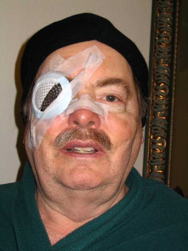
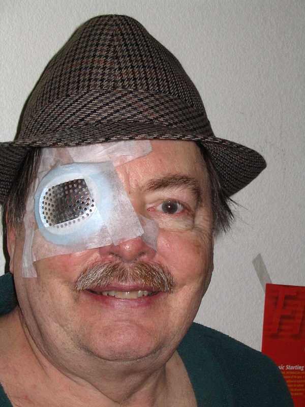
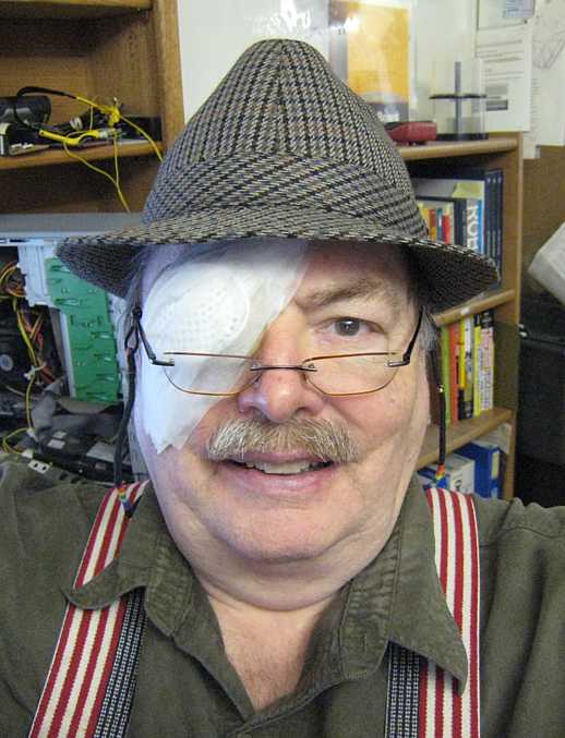
BillBill gave me that hat from Paris:-)) He wants to upgrade my image :-| Maybe I should also get my teeth whitened?
Days After - March 15, 2006
The day after, I went to the Dr.'s today to get that big patch off.
In preparation to see the doctor, a medical technician,
removed the patch to measure the eyeball pressure.
My first glance out was startling -
water hose size blood vessels on a field of straw yellow
The technician first wanted to check my vision
The technician (a real chick!) then put in some numbing drops.
About 20 minutes later Dr. Lehmer came in -
My eye will be teary and a little uncomfortable for a while
While at the pharmacy, I noticed an odd thing,
(Actually, Dr. Lehmer says my right eye will never be as good as my left eye
If you are into pharmaceuticals, here are the drops what I am taking: (text from internet)
Neomycin sulphate - bactericidal in action (kills bacteria). It does this by causing the bacteria to produce defective
proteins which are essential for their growth.
Polymyxin B sulphate - drug of choice in the treatment of infections of the urinary tract, meninges, and
blood stream, caused by susceptible strains of Ps. aeruginosa. It may also be used topically and subconjunctivally
in the treatment of infections of the eye caused by susceptible strains of Ps. aeruginosa.
The following few days
If I stay awake longer, the eye really tears up and feels scratchy and puffy -
I sleep with the patch on to reduce hazard.
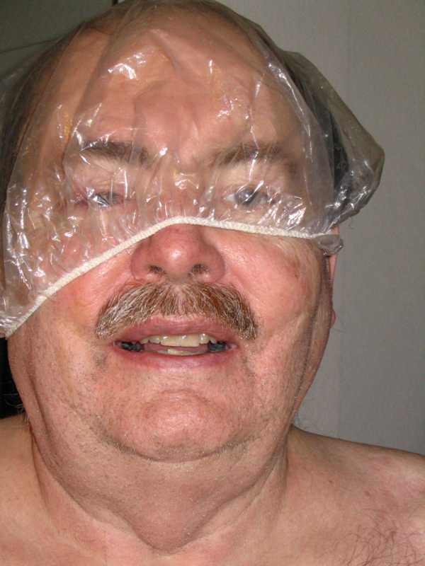
But Betty says I either shower daily or sleep on the floor - think, think, think - and look at this :-))
1 Week Checkup - March 20, 2006
It is now Monday - next check up - appointment at 3:15 :-|
I can definitely read the left letter below the big "E" on the eye chart. Its rather
wiggly - but definitely a "Z" :-)) I can only guess the other two letters. The technician numbs and dilates my eye, says my pressure is 17 (good).
Then I wait an hour. Dr Lehmer comes in but can't log in.
Gets a message with a big red "X". "What is this?"
He calls to the guy who has been walking up and down the hall for the past hour, who is apparently Software Services or something,
installing something. After a few rounds of experimenting, the software guy says "I don't know what is going on, but always
click "YES". and leaves.
I have an appointment for Thursday April 6th 3:15. Dr. Lehmer says I should be seeing even better then as my retina relaxes and flattens. :-))
2nd Week - March 27, 2006
I now appreciate their problem. - In a dark room, if there is a night light about 60 degrees to the left of where I
am looking, I see a thin ring of light about 30 degrees to the right of where I am looking. The length of the ring
is about +- 15 degrees about the axis of where I am looking.
Even in normal light, I get the feeling that an eye lash is hanging to the left of my field of view.
I am getting used to it - wonder if I should inform the doctor or if this is a standard "side effect"?
Having the mother of my children wander off turned out to be not much fun either.
So when I was diagnosed with macular degeneration (basically my right eye was on a one way trip to nowhere) I could almost
cheerfully say that it could be worse. Just hoped that the right eye would continue to track with the left eye so I wouldn't
bother others, and that the left eye would hang in there.
Since then, other doctors said the problem was "macular pucker" and could largely be corrected. Operation about 3 weeks ago, with
much progress since.
Woke up a few minutes ago, and noticed that the eye chart distortion could be described with a single pincushion distortion function.
The multiple crazy swirls had slowly become less, and were now almost nil.
As a test, I can play a solitaire using only my right eye and get the cards right with out too
much effort. I might not be legally blind in that eye - the state might let this old man drive for a few more years -
if my brain and other parts hang in there.
I thought of the old pictures of weeping women, with mouths agape, bodies and arms stretched forward, thanking a doctor
or other miracle worker for saving their child.
Boy - can I relate! - I'm sitting here with tears streaming, wishing to thank the doctor for giving me back my
now less screwed up eye.
I visit the Dr. for a checkup tomorrow (April 6) - hope I don't make an idiot of myself.
3 Week Check up - April 6, 2006
A bit of an anti-climax. Went to Kaiser for check up. Got numbing and dialating drops. Contact gauge said 14 mm mercury eyeball pressure.
Medical aid said that was in normal range - apparently 11 to 24? I could guess the second line of the eye chart better -
and even tried the third. Looking through the multi-pin hole mask helped even more. She then photographed my right eye with a machine showing
a cross of tiny laser looking dots.
Dr. Lehmer came in - looked in my eyes with another machine and a hand held light and with said things were coming along nicely. At my next
appointment, April 29th, he would fit me with glasses. We discussed eye drops, the 2 times a day drop, cut back to once a day, and all others
three times a day. I asked to get a copy of my macular angiogram, and was directed to the Medical Secretary to make the request. Should arrive by
mail in 2 weeks.
Next appointment April 29th, glasses??
6 Week Check up - April 29, 2006
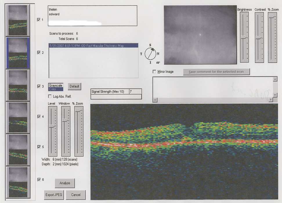
I showed up, got dialated, pressure checked (17mm), OCTed (a depth scan of the retinal area), and Dr. Lehmer came in. Apparently
there was still too much swelling at the macula to prescribe glasses. :-| We discussed the eye drops. Reviewed the frequency - to be the same,
except the antibiotic has run out - not to be renewed. A new appointment for Fri May 26th - at 8:00 in the morning - I thought only
bakers went to work that early - but the scheduler said that lots of folks are at work by then.
The requested angiogram has arrived. Look here.
Here is an e-mail dialog with my doctor - :-)) Sent via a log-in method.
If you are wanting the actual photos (as opposed to a written report based on the photos), then you may need to pay for a special photo processing fee to obtain them, but you should still be able to obtain them since they are a part of your record. You may print this e-mail out in order to present it to the person or persons you are speaking with regarding obtaining your records. I hope this helps.
The requested angiogram has arrived. Look here.
The right image shows the macula as the usual recessed region or dip in the average surface
Sorry about the break in reporting - The ability to see with my right eye reached a plateau.
Dr. Lehmer did not seem satisfied that the "macula" had reduced enough in swelling. So June 30th
he suggested two options:
The August 10th visit yielded
We discussed the likelihood of infection and other complications. I asked if it was his eye in this circumstance, would he choose
to have the injection. (I don't think he likes this kind of question.) He kind of mumbled that he thought so.
So - more drops and some antithetic swabbing of the eyelid area, he drew out some of the fluid that he had put in in March to make room for the
drug which he now put in.
After that procedure, I broke out into a cold sweat and felt light headed while sitting. We had a fun talk about my heart rate going
way down that being a more suitable reaction after a tiger tore off my arm. The lowered blood pressure would ease the bleeding,
and if I didn't get eaten, my chances for survival would be increased. We also discussed antithetics vs. "biting the bullet" of the Civil War,
- and - whiskey vs the current preoperative sedatives :-))
Soon I recovered and fled the area, after a good bye hand shake.
It is fun - the doctor thinks like I do - what is the Darwinian survival improvement of - you name it - skin color, hairy vs. little hair, ...
Well folks - I still can not read a book (or monitor with average font size) with my right eye - although I can now make out the
big E at the top of the chart, and fake it down a couple of lines. :-))
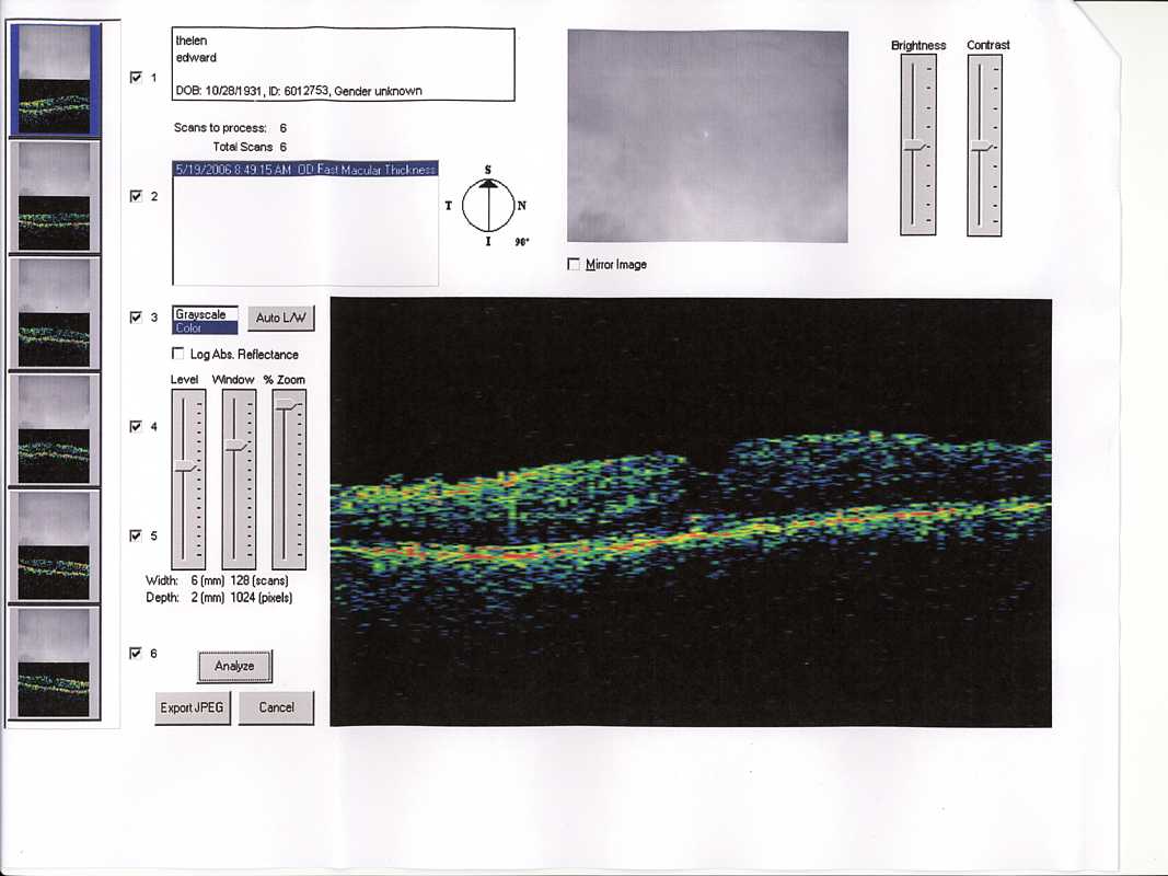
This is a late January OCT (Optical Coherence Tomography) scan.
Things of interest to Dr. Lehmer seemed to be:
Dr. Lehmer has tried all his tricks, and waited for effects. So he called for another Fluorescein Angiogram
which looked boringly similar to me but very interesting to him.
He said that likely the "internal limiting membrane" (separates the retina from the vitreous) under the previous scar tissue
has not flattened back to normal but has creased
crossway to the previous stress - He said that if he removed this membrane, the rest of the retina might revert more to its
previous unwrinkled state - He said the chances were good - and the risk no greater than the previous operation.
He reminds me that there has been some permanent damage to the sensor cells in the macula. (I believe this occured because
of Dr. Billie Knight's mistaken diagnosis of macular degeneration - and the delay permitted the increasing pucker to cramp the capillaries
reducing nutrients to the macula and harming the cells.)
Since the images from my right eye are more of a distraction than useful, and if something happens to my good left eye ...
OK - the stitch last year didn't feel scratchy all that much - just do a good job ;-))
Anesthesiologist
So March 1st 2007 I visited the Kaiser Hayward Anesthesiologist's office and three friendly crisp ladies, one a Nurse Practitioner and an
other an MD., gave me a going over - EKG and all - that I thought sufficient for a heart transplant.
My goodness - the game is that I will be sedated (and the eye anesthetized), but I am again to retain consciousness
so that I will not toss and roll my eyes at a bad time.
So, - Wednesday March 7, 2007 I have a preop with Dr. Lehmer,
Lovely Operation
It is the fateful day - March 8th, Thursday, I've had a lovely operation.
Dr. Lehmer had shown me a catalog from DORC (Dutch Ophthalmic Research Center)
- look for "Catalog", then "Vitreoretinal Instruments: ", then 25 gauge instruments.
He said that Bausch & Lomb Instrument Division also makes a line of similar instruments.
Cutie - really neat! - bags up the metal eye protector and suitable tape and says that I should sleep with the eye protector
taped on for the next week. Just think, a couple of generations ago I was so shy!!
The cutie leaves and I read my book. Dr. Lehmer comes in and looks through the usual (I have no idea what to call it) machine and seems
very satisfied. I ask about the fixed occlusions and he says that is ?kelvar? sticking to the retina, and will soon dissolve. He says
that things should be out of focus but improve over the coming weeks as the swelling of the retina goes down. He seems happy,
and if he is happy, I am delighted!
Dr. Lehmer then gives me a copy of the angiogram as I had expressed that I had been getting quite a run-around this time about getting a
copy. It seems that someplace else must be the correct place to get the pictures. Dr. Lehmer explains that I should advise the
Medical Secretary in Building A about his giving me the print, and gives me a note about what to say. I go the the Medical Secretary window,
(by now I could probably find it in the dark) and the person
there looks at the note and pictures and says that he knows how to pass requests to Doctors, and does not know what to do with this.
He asks another person in the office and that seems no help - and hands the material back to me. I feel that I have done my duty and exit stage left.
The above is the good news - now the problem -
Anyway, I read the warnings - and my eye feels so good - why risk a good thing. Still wondering if I should obey instructions
or "be safe". Really - a dilemma -
While writing this, I notice the occlusions noted above are no longer black, but light grey - but I cannot see through them to read.
1 week exam March 15, 2007
The pressures in the eyes are 16 and 17 mm, I forget which for which. The pressures are completely normal - OK.
The eye chart a little better (20-50?) but the funny black/grey stuff in/near the center of focus remains. Dr. Lehmer says they will go away slowly.
The Dr. says to stop taking drops from the little bottle, and use the big bottle - Polymyxin B sulphate - 3 times a day.
I Give Up - The End - May 1, 2007 - Ed Thelen - ed@ed-thelen.org
Well, I give up -
I have canceled the follow-up appointment for May 7, 2007 and am resigned to
my previous fate. Still having fun, hope the left eye hangs in there.
Signing off maintenance of this web page.
Cheers
2 Years Later
Stuart G. of Sherman Oaks, CA e-mailed wanting to talk on the phone - didn't think e-mail would do it for him.
Turns out he had the same thing (even a Kaiser surgeon and outside surgeon), same eye, same worries -
He pointed out that the above section sounded rather negative to the success of my operation.
Indeed, I was feeling somewhat negative at the time
Prologue
The subject - my right eye - is a big deal to me - I have only two eyes.
and my right eye couldn't resolve the big "E" at the top of the eye chart.
Most of the stuff (I hope) has happened,
I don't like mysteries, life is enough of a mystery, we hardly know what light is!!
Divergent Medical Opinion
A couple of years ago I went to an oculist at the local Walmart -
who informed me that my right eye was really bad - that I might have "macular degeneration" :-((
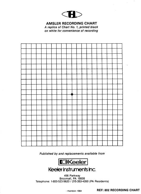
My left eye saw this as regular graph paper with a dot in the center.
My right eye saw this as wildly wiggly lines, from 16 inches away, near the center of vision,
the wiggles were about 1 or more units wide and about 2 or 3 units long. There were wiggles
that spread black lines wider and narrower, and wiggles that looked like pincushion camera distortion.
When you try to draw it, you move your eyes and the wiggles all shifted around :-((
But macular pucker is reversable - the eye can be useable again - but likely not 20-20, hopefully 20-30. :-))
Concurrence
Dr. Lehmer
(same spelling as the family of
Berkeley mathematicians, but nothing close) said that the
scar tissue was crumpling the back of my eye by pulling it together into little mounds and folds. He said that
this often interferes with blood flow, and can cause its own form of macular degeneration due to lack of
nourishment and oxygen from the restricted blood flow. He ordered an angiogram - I thought angiograms were of the heart,
but in his business it checks blood flow in the back of the eye.
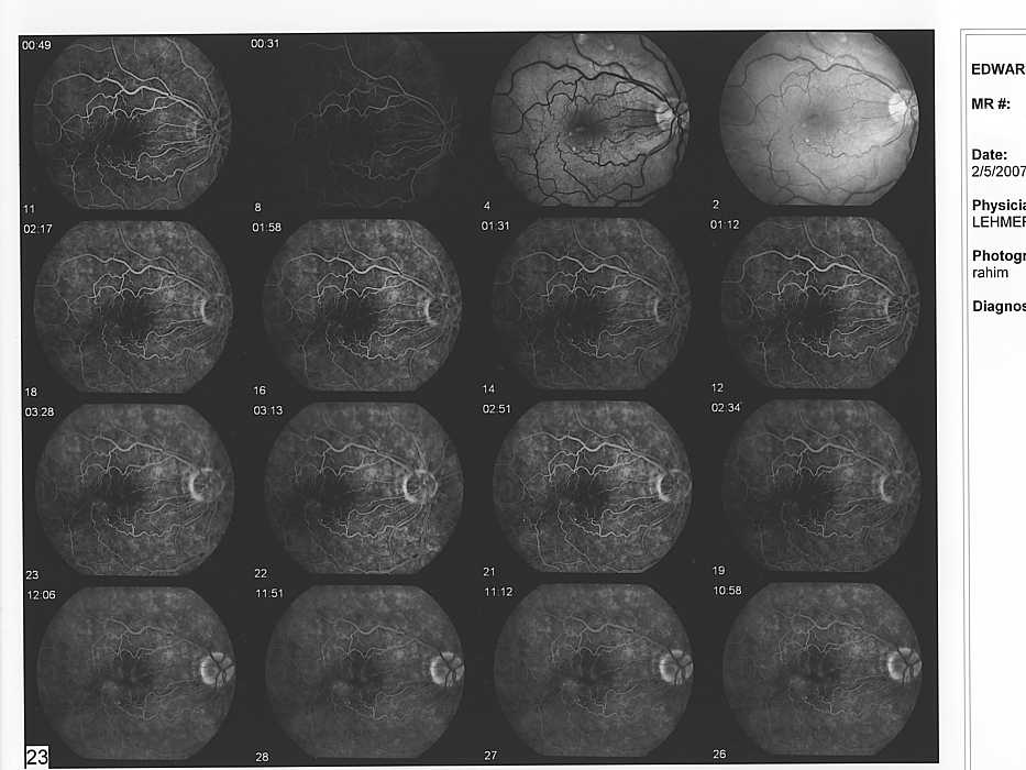
Fluorescein Angiogram Torture
2006 vs 2007
A few days later I show up for the Fluorescein Angiogram. Careful asking yielded that they would inject the dye into
a vein?/artery? on the back of my hand - just relax. OK. The eye drops of course, and the hypodermic are inserted (no dye yet).
Just watch the little green dot - lets take some before shots - **DAMNIT THAT LIGHT IS BRIGHT** HOLLY BALLS!!
Good News
Monday morning the next week Dr. Lehmer calls me and says he has some good news - in a not good news voice.
I wait for the "and some bad news." But there is no bad news - the back of my eye is good enough, he offers to operate on me.
I still wait for the bad news - no bad news. He offers two dates the closest is next week Tuesday the 14th.
We go over again the failure modes and percentage expectations again. The kicker in the background is serious
infection that will ruin/destroy the eye - less 1/300, and the Internet quoted even less. Of course, these
guys make a living selling "procedures", sound too bad, scare the patient, and you are out of business - interesting -
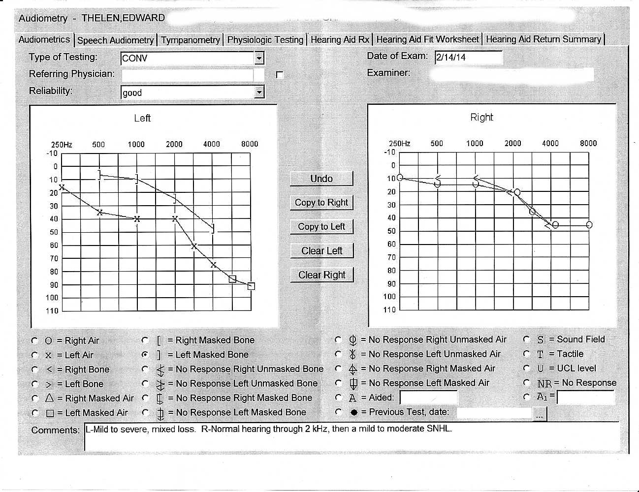
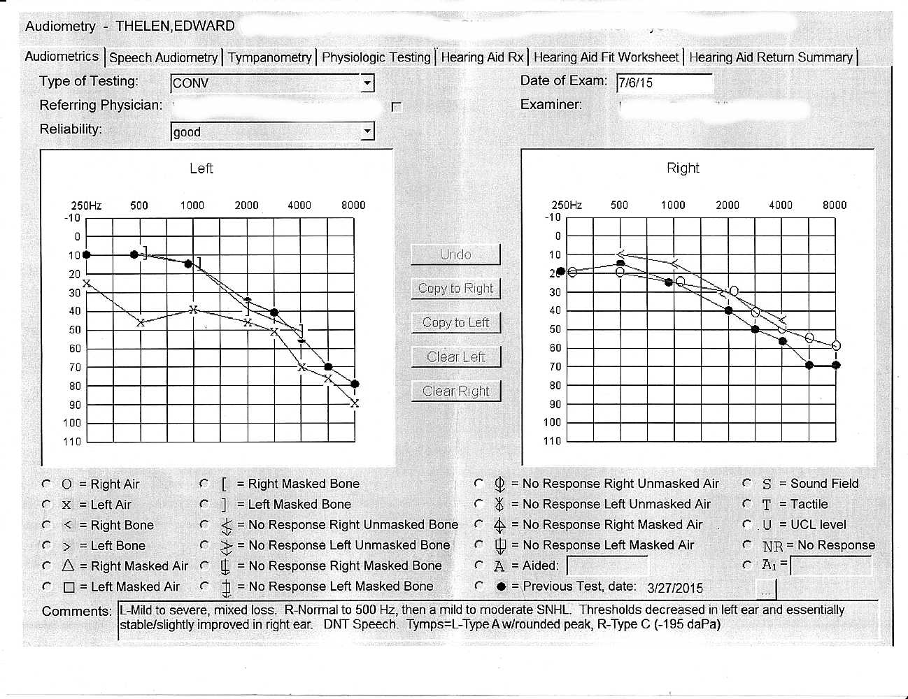
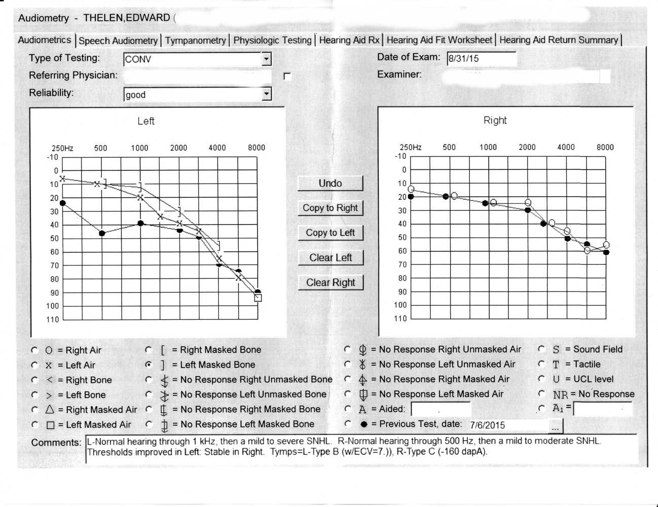
Left eye
Right eye
Comments
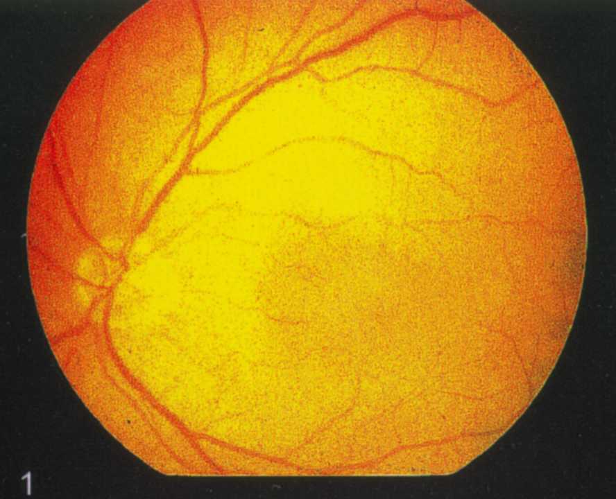
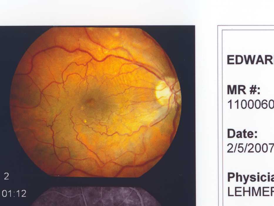
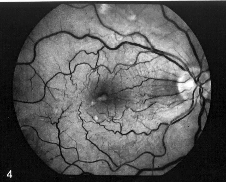
Color - before
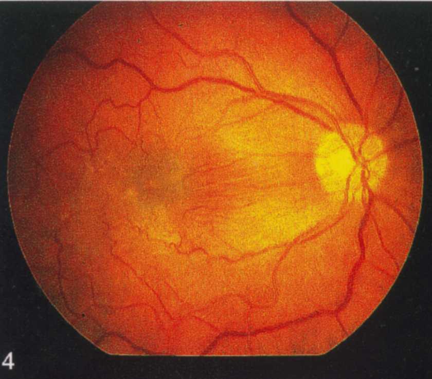
gray scale - before - The dark area near the center is the macula - fovia
.

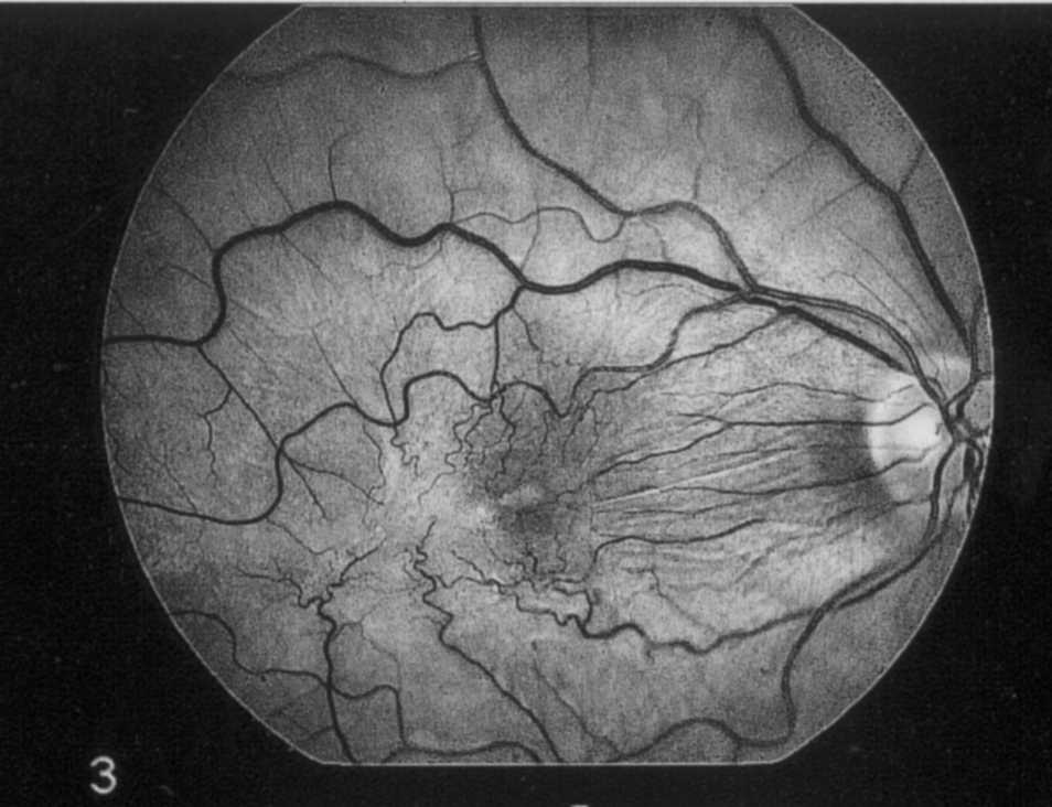
gray scale #05 - about 20 seconds after the fluorescein dye was injected into the back of my hand
.
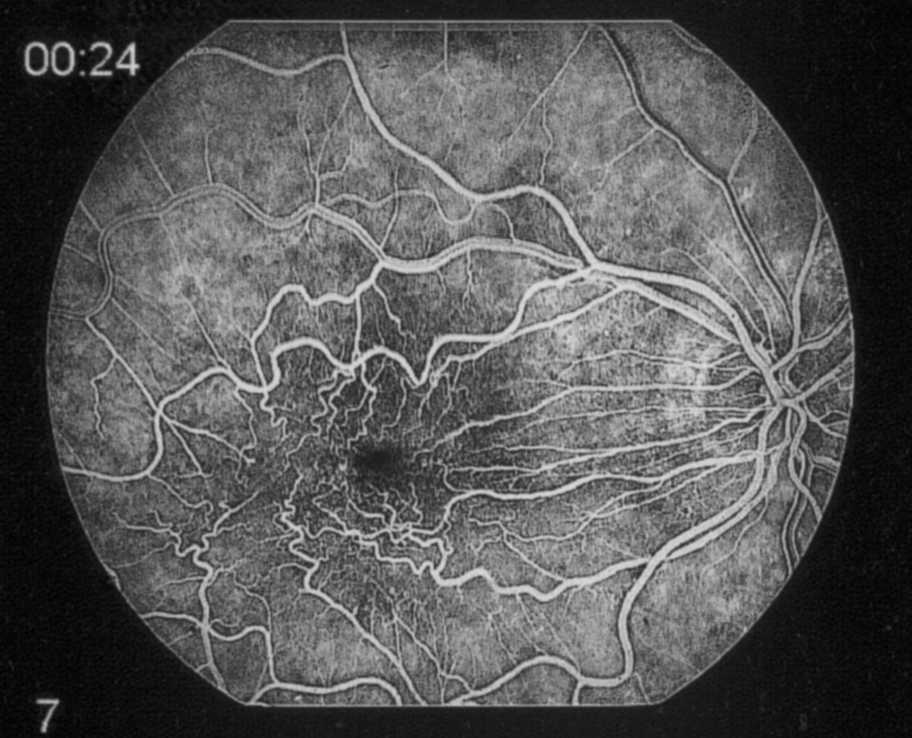
gray scale #07 - after
.
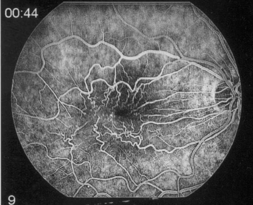
gray scale #09 - after
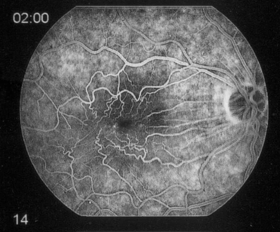
gray scale #14 - after
the left image is my left eye about the same time
.
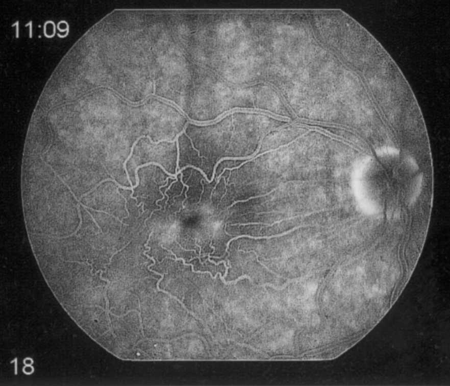
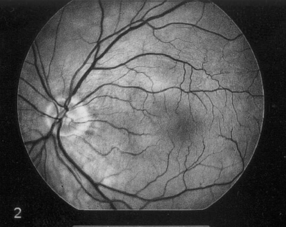
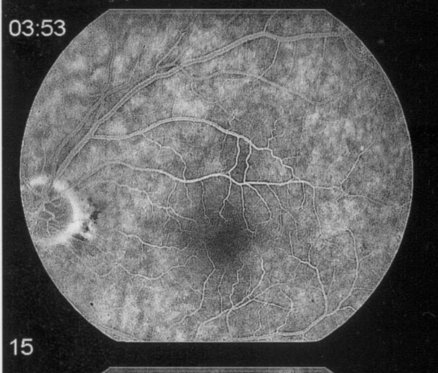
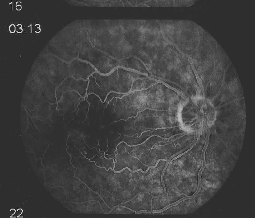
gray scale #16 - After a ?15? minute wait for any leakage to show.
The gray areas near the black macula indicates leakage of the fluorescein
dye from the strained capillaries.
that I signed has - as best I can decipher:
(the medical assistant's writing is big and round, and when squeezed, as hard to read
as we tease doctors about)
The Night Before
So - it is midnight and I have quit eating, drinking,
cussing, and other bad habits six hours ago :-((
is given
and that I will be "fully awake".
I just want to die in peace :-|
take the following:
I REALLY hope I sleep for the full 100+ minutes,
OH GOD !!
OH GOD!!
If I had wanted to run, I might as well be wearing prison strips.
Well, I died a few more today laying all preped up
for 1 hour, 25 minutes staring at the ceiling
wondering if all this was worth it.
Various people are wandering in and out
It is much cooler in here.
Jake the nurse anesthesiologist is here
Things must be OK if they are making chitchat.
Then a voice says "OK, All set, Just fine".
Betty goes to get the car,

Day00-Tuesday. I have survived. The anesthetic is still active. The mild scratchy feeling
has not started. (The doctor said that if there was pain to take a non-aspirin pain killer -
there was never any pain. But that eye sure wept a lot - especially if I got tired.
I take three types of eye drops (Betty does it.) Four, three, and two times a day. Schedule that!
What the heck
Within 30 seconds, this view was fading out,
The chick technician wrote this down, and left -
That makes three eyedrop medicines with deliveries,
It hit me - THE OPERATION WAS A SUCCESS !!!!!!!!
Prednisolone acetate (microfine suspension) 1% optht drops
4 times a day
1 refill
Topical steroids - For moderate to severe inflammation - Because it is an acetate suspension, prednisolone acetate
is optimally suited for penetrating the cornea and the anterior chamber. This makes it effective for treating anterior
chamber inflammation and remains the drug of choice in treating anterior uveitis.
Dexamethasone/neomycin sulphate/polymyxin B sulphate optht drops
3 times a day
0 refills
Dexamethasone- a corticosteroid, is similar to a natural hormone produced by your adrenal glands.
It often is used to replace this chemical when your body does not make enough of it. It relieves inflammation
(swelling, heat, redness, and pain) and is used to treat certain forms of arthritis; skin, blood, kidney, eye,
thyroid, and intestinal disorders (e.g., colitis); severe allergies; and asthma.
Timolol Maleate 0.5% optht drops
2 times a day
3 refills
(This was prescribed the day after the "procedure" when my eye pressure was 26 somethings rather than 18 somethings -
non-selective beta-adrenergic receptor blocking agent. Timolol maleate, when applied topically on the eye,
has the action of reducing elevated as well as normal intraocular pressure, whether or not accompanied by glaucoma.
Elevated intraocular pressure is a major risk factor in the pathogenesis of glaucomatous visual field loss.
The higher the level of intraocular pressure, the greater the likelihood of glaucomatous visual field loss and optic nerve damage.
Day02-Thursday - I am sleeping 15 hours a day - basically sleep 5 hours, awake 3 hours, ?like a baby?
Day03-Friday - All the medical people stressed keeping shampoo and water out of my eye.
Day05-Sunday - I was up for 12 hours in a row Saturday. Betty wanted to go to some shindig in San Mateo Saturday night,
and wanted me to drive.
BUT - it is evident that there is some distortion remaining.
Betty and I can't figure what that orange flare in my right eye is. The pupil is still a little dilated, but both eyes and camera see it.

Day06-Monday - Dr. Lehmer examines my dilated eye - and says "Good".
Well - Monday again - getting lots of sleep - all seems getting better - can see things like many of the vertical fins on a wall ventilator
- the rest blur -
Still taking all the eye drops (thanks Betty), and wearing the protective patch at night - I don't want to screw up!
Two things seem interesting:
The friend of mine who had both lenses replaced had mentioned that he finds driving at night difficult and distracting.
I will ask about his "internal reflections".
Pathetically Grateful
I've always figured the worst thing would be to lose a child, or drive along and have someone else's child dart in
front of my car.
From: THELEN,EDWARD S
Sent: 5/2/2006 12:52 PM
To: Office of JEFFREY MARK LEHMER MD
Subject: unable to get copy of "angiogram" of my eye
On April 6th I requested that I get the Laser
fluorescent image of the macular region of my eyes.
This seemed to be called an "angiogram" even though it
did not involve X-rays and the heart.
The medical secretary (MaryAnn 510-675-xxxx) says
that "angiogram" is not the correct word - but in any
case there are no round gray scale images of my retina
in my file. She does have the OCT of Jan 13th, but
nothing more recent.
What can I do? What should I call it? Help :-))
To: Edward S Thelen
From: JEFFREY MARK LEHMER MD
Subject: RE: unable to get copy of "angiogram" of my eye
Received: 5/2/06 3:33 PM
Dear Mr. Thelen:
The study you are asking about is called a Fluorescein Angiogram of the Retina. It does NOT involve laser or x-rays. It involves the injection of a fluorescent yellow vegetable dye called fluorescein into the arm vein followed by flash pictures of the retina over a 10 minute time sequence using a digital camera called a retinal fundus camera that uses special filters to capture the fluorescence of the dye as it passes through the retinal blood vessels.
Sincerely,
Jeffrey Mark Lehmer, M.D.
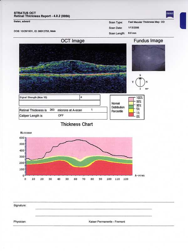
Pre operative OCT scan
Post operative OCT scan
The left image is preoperative. I am guessing that the black line in the thickness chart is my macular thickness,
and the green zone in the usual.
August 10th Check Up
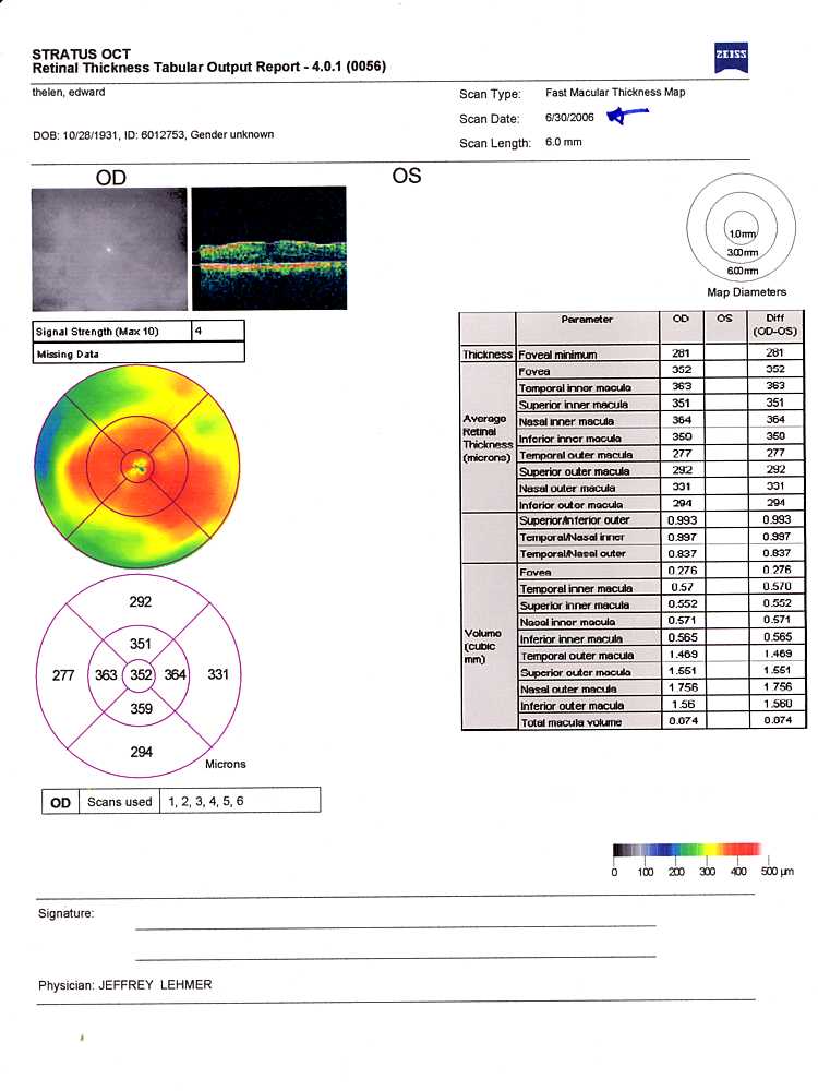
June30-OCT - Dr. Lehmer wishes to get the number in the center bulls eye down toward 250 (nanometers) (typical) from the 352 shown. He also
pointed out the little black spaces near the macula as suspected pockets of fluid - apparently contributing to the thickness and swelling?
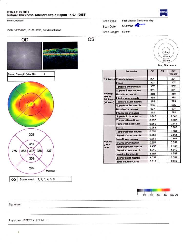
August10-OCT- a little better - 337 down from the previous 352 - but it is so slow that I accepted his suggestion
of a shot of cortisone or steroid or ... in the right eye -
which is likely to speed reduction of swelling.
March 2007 - Lets try again
The thickness of the retina (red) of my right eye
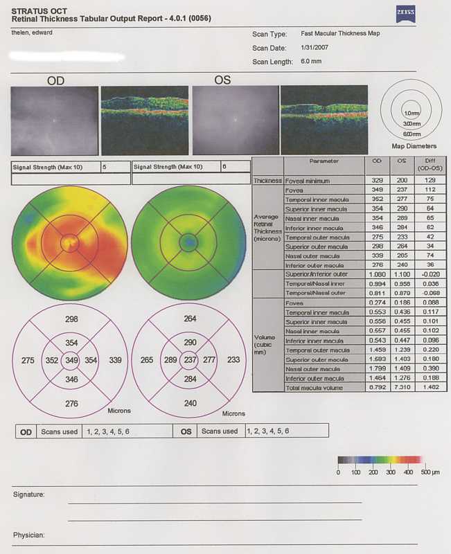
The flatness of the area to the right of the macula/fovea (the little dip)
He said:
Even though he already had me sold, he continued on, saying that the technology had improved since last year. This year he would
be operating through a # 25 needle, instead of the thicker # 20 needle of last year. Since I don't like needles of any size,
I get queazy, until he said that the needle is so thin that they don't need to even take the one stitch of last year.
The hole is so small that the eye white membrane would just close it over - similar to taking a blood sample. And the patch can come
off after a day instead of a week :-))
Dr. Lehmer shows me a catalog from DORC (Dutch Ophthalmic Research Center)
- look for "Catalog", then "Vitreoretinal Instruments: ", then 25 gauge instruments.
(With the # 20 needle last year, and the stitch, the patch came off after a week.)
I ask the doctor if he likes using the # 25 needles, I would rather have him use familiar tools rather than just not take a stitch
and its longer recovery. Dr. Lehmer says that he prefers the # 25 equipment - that he uses this in his practice and has been try
to get Kaiser to adopt such. He says an additional advantage is that if there is a problem - (eye movement or such?) -
the smaller equipment will do less damage. (I like folks that can talk straight up about risks and problems.)
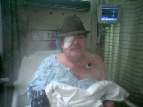
It is now 6 hours later and still comfortable - and hopeful ;-))
A nice time in the recovery area,
- Please excuse the cell phone camera image :-((
And very comfortable at home with out even an aspirin. No apparent worry about a scratchy feeling from the stitch that wasn't
needed with the # 25 instruments.
Now there is something fixed long enough to describe and roughly draw - well a change anyway - and I think the wiggles
are mostly? gone? Anyway, if you are focusing on the middle letter of this "eye chart", this is roughly what you see.
The right hand side of what you are looking at is obstructed by that black funny thing, and to the lower left is another odd thing.


This is "the eye" one day after the second operation -
so much better than 1 week after the first operation. Of course, this time the lens wasn't jellyfied and removed
and a rolled up plastic one inserted.
see descriptions above
Cheers,